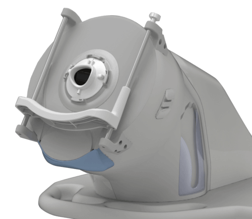
Very High Frequency Robotic Ultrasound provides imaging of the anterior segment anatomy and pathology of the eye. With multiple preconfigured Scan Types to select from, diagnostically significant information can be gleaned from the precise detail not visible through optical scanning methods. Detailed biometric information includes corneal layers, and anterior and posterior chambers, offering repeatable visualization of the eye that can support surgical and treatment planning.
Each category below leads to specific scan and the relevant measures clinicians may create. With the flexibility of the manual scan (Scout Scan) option and the power of image review, the operator adds their expertise to the process too.

Scan Set Applications
How Scanning Works
Each Scan Set was programmed based on a specific focal plane, number, and location of meridians, delivering key data for treatment and surgical planning. The software captures 2mm of data in front of and behind the focal line, captured in an arc to match the cornea’s radius of curvature.
With careful positioning of the patient, the device scans in an arc to maximize the angle an depth of the ultrasound. The multiple scans that make up each image are efficiently captured with the robotically guided transducer probe. From start to finish, the patient is comfortably seated for under 10 minutes, with experienced operators dramatically reducing that time.
